College of Liberal Arts & Sciences
6C20.18 - Interference Patterns - Water Drop

Code Number:
References:
6C20.18
Demo Title:
Interference Patterns - Water Drop
Condition:
Good
Principle:
Interference of Reflected Light
Area of Study:
Optics
Equipment:
5 cc to 60 cc Disposable Syringe, 14; 15; 16; or 18 gauge long, blunt Needles, 5 mw Laser, White Screen, Water.
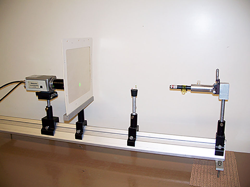
Procedure:
A long, blunt 16 gauge needle seems to work best. Put the needle on the syringe and clamp in a vertical position so that you can hang a drop from the end of the needle. Shoot the laser at the hanging drop at a slight downward or horizontal angle which seems to work best. Try to aim the laser into the middle of the drop so that the patterns can be seen as you move the laser from side to side (The needle end provides an interference of its own if you try to do this by moving the laser up and down). The interference pattern will be reflected back to, and to the left or the right of the laser. A white screen may used to make the pattern image sharper. The interference pattern is produced by reflections from the front and back surfaces of the hanging drop. Due to the distance between the surfaces and the curvature of the surfaces the pattern is produced.
An interesting side effect is that microscopic particles and contaminants in the drop are also visible. They show up as a floating, circular interference pattern, that looks much like the Michelson interference pattern that is shown in 6D40.10. The curved surface of the drop acts as a magnifying lens.
- Mark Welter, "Interference of Laser Light Using a Lab Beaker", TPT, Vol. 54, #8, Nov. 2016, p. 505.
- Gorazd Planinsic, "Water-Drop Projector", TPT, Vol. 39, #2, Feb. 2001, p. 76.
- David Keeports, "Estimating the Size of Blood Cells by Staring Into Space", TPT, Vol. 36, #1, Jan. 1998, p. 58.
- G.R. Davies, "Interference and Diffraction Corridor Demonstrations", TPT, Vol. 33, #4, Apr. 1995, p. 244.
- George W. Goth, "Measuring the Sizes of Biological Samples by Diffraction", TPT, Vol. 33, #9, Dec. 1995, p. 556.
- David R. Lapp and James E. Keenan, "Cow's Eye Dissection in the Physics Lab", TPT, Vol. 29, #8, Nov. 1991, p. 502.
- R.D. Edge, "The Optics of the Eye Lens", TPT, Vol. 27, #5, May 1989, p. 392.
- Y. P. Hwu, "Diffraction Pattern of Blood Corpuscles", TPT, Vol. 17, #4, Apr. 1979, p. 258.
- Dr. J. E. Parks, "If Dracula Had A Laser", TPT, Vol. 17, #5, May 1979, p. Back Cover.
- L. Orwig and G. Schrank, "Simple Laser Scattering Experiment for Biology-Oriented Physics Labs", AJP, Vol. 47, #6, June 1979, p. 500.
- Jearl Walker, "Floaters": Visual Artifacts That Result From Blood Cells in Front of the Retina", The Amateur Scientist, Apr. 1982.
- Jearl Walker, "7.5 Floaters and Other Spots in Your Eye", The Flying Circus of Physics Ed. 2, p. 244.
- T. D. Rossing and C. J. Chiaverina, "#2, "Floaters" In The Eye", Light Science, Physics and Visual Arts, p. 125.
- "Waterdrop Microscope", Laser Classroom.
Disclaimer: These demonstrations are provided only for illustrative use by persons affiliated with The University of Iowa and only under the direction of a trained instructor or physicist. The University of Iowa is not responsible for demonstrations performed by those using their own equipment or who choose to use this reference material for their own purpose. The demonstrations included here are within the public domain and can be found in materials contained in libraries, bookstores, and through electronic sources. Performing all or any portion of any of these demonstrations, with or without revisions not depicted here entails inherent risks. These risks include, without limitation, bodily injury (and possibly death), including risks to health that may be temporary or permanent and that may exacerbate a pre-existing medical condition; and property loss or damage. Anyone performing any part of these demonstrations, even with revisions, knowingly and voluntarily assumes all risks associated with them.